Imaging systems have become a cornerstone of modern healthcare, playing a critical role in diagnosis, treatment, and ongoing patient care. These technologies allow physicians to view the human body in detailed ways that were unimaginable decades ago, enabling them to diagnose diseases accurately, monitor treatment progress, and predict health outcomes more effectively.
Medical imaging has progressed significantly over the years. According to a paper published on NCBI, imaging informaticians are now crucial players in implementing AI in the medical setting, indicating how technology is revolutionizing this field. Another article highlights the importance of improving access to medical imaging, noting that a lack of access can dramatically affect health outcomes.
The World Health Organization (WHO) notes that diagnostic imaging services are essential in confirming, assessing, and documenting the course of many diseases and their responses to treatment. According to an article on Medium, these services are crucial to the American healthcare system.
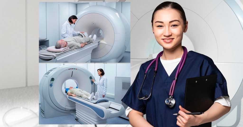
Introduction to SPECT and PET Imaging Technologies
Among the various imaging technologies available today, Single Photon Emission Computed Tomography (SPECT) and Positron Emission Tomography (PET) stand out due to their unique capabilities and applications.
Single Photon Emission Computed Tomography (SPECT)
As one of the most commonly used nuclear imaging procedures in healthcare, Single Photon Emission Computed Tomography (SPECT) plays a vital role in diagnosing and monitoring various conditions. It produces three-dimensional (3D) images of the body’s organs using a radioactive material and a specialized camera.
Role and Benefits in Healthcare
SPECT is beneficial in numerous ways. For instance, it helps detect areas of the heart that have low blood flow during rest or exercise, providing valuable information for healthcare professionals. This non-invasive nuclear imaging technique is also used to determine how internal organs function, offering insights that can guide treatment decisions.
Furthermore, the advent of SPECT-CT, which combines SPECT with computed tomography (CT), has improved patient outcomes by providing more detailed images. These hybrid imaging systems have been commercially available since the early 2000s.
Specialization and Applications
SPECT is primarily used in cardiology and neurology. In cardiology, it’s employed to diagnose heart conditions like coronary artery disease, while in neurology, it’s used to investigate conditions such as dementia, epilepsy, and stroke.
Origin and History
The concept of SPECT originated in the early 1960s, but it wasn’t until the late 1970s that the first commercial SPECT systems became available. Since then, the technology has evolved significantly, with over 18 million SPECT procedures now performed annually in the healthcare system.
Positron Emission Tomography (PET)
Positron Emission Tomography, commonly known as PET, is another crucial technology in the field of nuclear medicine imaging. It uses a small amount of radioactive material, a special camera, and a computer to evaluate organ and tissue functions in the body.
Role and Benefits in Healthcare
PET scans are instrumental in several areas of healthcare. They are used to detect cancer, evaluate brain disorders, and identify heart disease. PET scans can show how tissues and organs are functioning, making them especially useful in detecting cancerous cells that tend to be more active than normal cells.
Additionally, PET scans are often used in conjunction with CT or MRI scans to provide more detailed information about where the abnormal cells are located and whether they are benign or malignant.
Specialization and Applications
PET scans are particularly useful in oncology for diagnosing and staging cancer, evaluating the effectiveness of treatment, and determining whether cancer has returned after treatment.
In neurology, PET scans can help diagnose conditions like Alzheimer’s disease, Parkinson’s disease, and epilepsy by showing areas of the brain that are not functioning normally. In cardiology, they can help identify areas of the heart muscle that may benefit from procedures such as angioplasty or heart surgery.
Origin and History
The concept of PET scanning was first proposed in 1953, but the first commercial scanners were not available until the early 1970s. Since then, the technology has evolved significantly, with advancements in scanner technology, radiochemistry, and image reconstruction methods leading to improved image quality and diagnostic accuracy.
Comparing SPECT and PET
While both SPECT and PET are nuclear imaging technologies, they differ in several ways regarding their uses, effectiveness, and specific applications. Understanding these differences is crucial for healthcare professionals as they determine the most appropriate imaging technique for a given patient or condition.
Uses
Both SPECT and PET are used to produce three-dimensional images of the body, but they focus on different aspects. SPECT primarily measures the distribution of radioactive tracer material in the body to give a snapshot of blood flow or organ function at a particular moment.
On the other hand, PET measures the metabolic activity of cells by detecting the radiation emitted by a radioactive tracer. This allows physicians to see how tissues and organs are functioning, which is particularly helpful in detecting cancerous cells.
Effectiveness
In terms of resolution, PET scans generally provide higher-resolution images than SPECT scans. This increased resolution can provide more detailed information about the presence and location of disease, potentially leading to more accurate diagnoses.
However, SPECT scans remain a valuable tool in many areas of medicine due to their lower cost and wider availability. For instance, in cardiology, SPECT imaging remains the standard for assessing myocardial perfusion, despite the higher resolution of PET imaging.
Distinctions
A key distinction between SPECT and PET lies in the type of radiation they use. SPECT uses gamma rays, while PET uses positrons, which interact with electrons in the body to produce gamma rays. This fundamental difference affects the types of tracers used, the cost, and the logistical requirements of each scan.
In terms of specialization, SPECT is commonly used in cardiology and neurology, while PET is widely used in oncology, neurology, and cardiology.
The Critical Role of Imaging Systems in Healthcare
The importance of SPECT and PET imaging systems in healthcare cannot be overstated. These advanced diagnostic tools have revolutionized the way illnesses are diagnosed and monitored, providing physicians with invaluable insights into the inner workings of the human body.
By producing detailed, three-dimensional images of organs and tissues, SPECT and PET scans allow for more accurate diagnoses, enabling healthcare professionals to target treatment more effectively. Whether it’s identifying areas of reduced blood flow in the heart, detecting abnormal brain activity, or pinpointing the location of cancerous cells, these imaging systems play a pivotal role in patient care.
While each system has its own unique strengths and applications, their common goal is clear: to provide the most precise and comprehensive information possible to guide medical decision-making. In this era of personalized medicine, the ability to visualize the exact nature and extent of disease within the body is invaluable.
As technology continues to advance, we can expect even greater strides in nuclear imaging, further enhancing our ability to diagnose and treat a wide range of conditions. Ultimately, SPECT and PET scans represent essential tools in the ongoing quest to improve health outcomes and quality of life for patients worldwide.